"Cardiac imaging tests are not just pictures—they can be lifesaving," said St. Francis Hospital & Heart Center® Director of Cardiovascular Imaging Omar Khalique, MD. "That's why, at St. Francis Heart Center’s Division of Cardiovascular Imaging, we use the most advanced technology that images the heart quickly and with great detail. This leads to more accurate diagnoses, allowing doctors to create customized treatment plans for their patients."
At your annual exam with your primary care physician (PCP), your blood pressure and heart rate are taken for a basic look at your general heart health. But your PCP may refer you for additional cardiac tests if you have:
- A family history of heart disease
- Repeated high blood pressure readings
- High cholesterol
- Frequently experience heart flutters, shortness of breath or extreme fatigue
- Been previously diagnosed with a heart condition or experienced a cardiac episode like a heart attack
Commonly assessed conditions include:
- Atrial fibrillation (AFib)
- Cardiomyopathy
- Congenital heart disease
- Congestive heart failure
- Coronary artery disease
- Mitral valve disease
- Pericardium disease
Cardiac imaging allows both a PCP and a cardiologist to accurately diagnose and prescribe treatment.
How Heart Healthy Are Your Habits?
Our quiz helps you make lifestyle choices that improve your heart health.
Echocardiogram (Echo)
An echocardiogram, also referred to as echocardiography, cardiac echo or echo, uses ultrasound (or sound waves) to create images of your heart. There is no radiation. A technician places a wand on your chest to take images of your heart, which are more detailed than standard X-ray images.
An echo lets your physician see how blood moves through your heart and how your heart is beating.
You may have an echocardiogram to:
- Assess your heart’s overall condition
- Check your heart valves
- Detect swelling around the heart
- Find the cause of a heart murmur
The different types of echocardiography include:
- Transthoracic echo—most common type of echo
- Stress echo—combines ultrasound with a stress test
- Transesophageal echo—uses a probe inserted into the mouth and down the esophagus
- Interventional echo—guides valvular and other percutaneous procedures
- Intraoperative echo—assists a surgical team with surgery planning
It takes about an hour to complete the test.
Electrocardiogram (ECG or EKG)
An electrocardiogram measures electrical activity and shows how well your heart is beating. Your physician uses an EKG to determine if your heart’s electrical activity is normal and if parts of your heart are too large or overworked.
During the test, a technician places small, sticky dots on your arms, chest and legs and attaches them to a machine that records the heart’s electrical impulses. It is not painful and does not send electricity into your body.
Your physician may recommend an EKG to:
- Check your heart’s rhythm
- Diagnose a heart attack
- Identify structural abnormalities within the heart
The test takes a few minutes to conduct.
Cardiac magnetic resonance imaging (MRI)
A cardiac MRI takes high-speed and high-resolution images of your beating heart that accurately illustrate the heart chambers, heart muscle, wall motion, ejection fraction and blood flow.
If your physician wants to see how well blood flows through your vessels, they will inject a special dye into your body before creating the images. This procedure is called magnetic resonance angiography (MRA). The contrast can also assist in Identifying abnormalities within the heart muscle.
You may have the test to:
- Assess your heart’s structure including the heart muscle and pericardium (a membrane, or sac, that surrounds your heart)
- Diagnose coronary artery disease
- Diagnose congenital heart defects
- Identify scar tissue in the heart
A cardiac MRI is noninvasive, does not use radiation and takes about an hour. With some exceptions, it is safe for those with cardiac stents, artificial valves, pacemakers and metal implants.
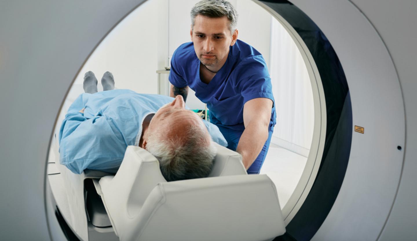
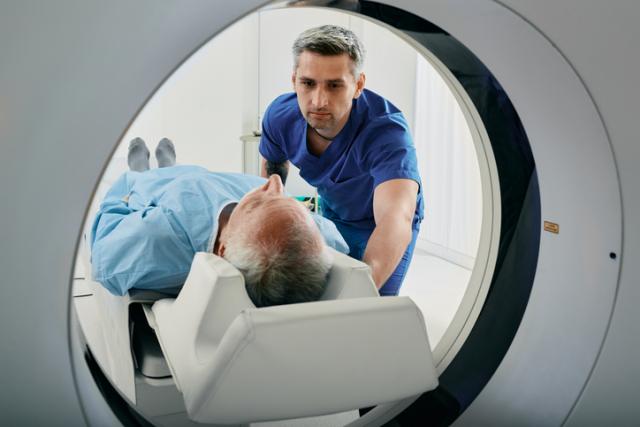
Cardiac computerized tomography (CT) scan
A cardiac CT scan is an X-ray that uses a computer to make 3D, motion-free images of the beating heart. It is also called cardiac computed tomography, computerized axial tomography or CAT scan. Physicians use this test to look for problems with your heart and blood vessels.
Cardiac CT scans can:
- Assess the structure of the heart
- Find blockages in the coronary arteries
- Identify disease in the membrane surrounding your heart
Your physician may also recommend a computerized tomographic angiography (CTA) that combines CT technology with an injection of a contrast dye to create detailed images of the blood vessels.
The test takes about 30 minutes.
Exercise stress test
An exercise stress test tracks your heart rate, breathing, blood pressure and heart activity during exercise. You’ll ride a stationary bike or run on a treadmill while an EKG monitors the blood flow to your heart. You will jog for 30 minutes to an hour. The results will help your physician:
- Assess your heart’s health
- Determine if it is safe for you to exercise
- Identify inadequate blood flow to the heart during exercise
- Locate the source of chest pain, shortness of breath or weakness
- Track changes in heart rhythm during activity
Cardiac nuclear imaging
Also referred to as a chemical stress test, nuclear imaging is helpful for patients who cannot complete simple stress tests that involve exercise. For this test, your physician will give you medication through an IV line in your arm. The medicine increases your heart rate and blood flow, mimicking the effects of exercise, while in a controlled environment.
The addition of nuclear imaging increases the diagnostic accuracy of the stress test, especially when coronary artery disease is suspected. After you receive the medicine, your physician will take images of your heart using ultrasound or a specialized camera. The images will estimate how blood flows to the heart.
A chemical stress test can:
- Determine the cause of chest pain, shortness of breath or weakness
- Estimate the risk of a heart attack
- Identify inadequate blood flow to the heart during physical activity
- Monitor or diagnose blockages in the coronary arteries
The test takes several hours to complete.
Tilt test
A tilt test lets your physician monitor your heart’s activity when you are lying down and standing up. Your physician may order this test if you report feeling faint or lightheaded.
During the test, you’ll lie strapped to a table that is slowly tilted upward to a standing position. Then, a nurse or technician tracks your heart rate and blood pressure to see how they respond to the different positions.
A tilt table test can help your physician:
- Detect changes in heart rhythm
- Identify sudden drops in blood pressure with position changes
- Pinpoint the cause of dizziness or fainting spells
The test takes about 90 minutes to complete.
Ambulatory monitoring tests
Holter monitoring (a wearable device), event recorders and mobile cardiac telemetry (MCT) study your heart rhythm while you go about your daily activities at home or work.
You’ll wear a small, battery-powered device that records your heart’s activity. Your physician will request you wear the monitor for at least 24 hours or up to several weeks. The device records heart symptoms that come and go.
Ambulatory monitoring tests can:
- Identify the cause of heart palpitations or symptoms
- Track heart rate and rhythm for an extended period
Coronary angiogram
Coronary angiogram, also called cardiac catheterization, is a test that uses a specialized X-ray to examine your coronary arteries. Your physician inserts a small tube, called a catheter, into a blood vessel in your arm or groin.
The tip of the tube is fed all the way to the heart and coronary arteries, where dye is injected. Next, a technician takes X-rays of the dye traveling through the heart’s chambers and valves. These images are called angiograms and help diagnose coronary artery disease.
Angiograms help your physician:
- Assess the size of your heart chambers
- Determine how well your heart valves open and close
- Identify narrowed or blocked arteries
- Measure pressure within the heart chambers and blood vessels
The test takes between 30 minutes and two hours.
Find care at St. Francis Heart Center
Catholic Health's St. Francis Heart Center is Long Island’s most awarded heart program. Our nationally recognized experts offer patients the latest, most advanced technology for cardiac imaging.
- St. Francis Hospital & Heart Center® (Roslyn, NY)
- St. Francis Heart Center at Good Samaritan University Hospital (West Islip, NY)
- St. Francis Heart Center at Mercy Hospital (Rockville Centre, NY)
- St. Francis Heart Center at St. Catherine of Siena Hospital (Smithtown, NY)
- St. Francis Heart Center at St. Joseph Hospital (Bethpage, NY)
Learn more about our cardiac imaging services.
Call 866-MY-LI-DOC (866-695-4362) to find a Catholic Health physician near you.